
Mole Mapping
*** Please note - All appointments are to be booked at The Private Medical Group's website:
www.theprivatemedicalgroup.co.uk
Mole Mapping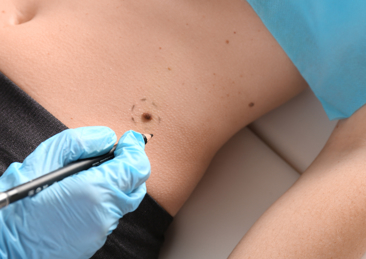
Early detection is key when it comes to preventing and managing skin cancer. Our Mole Mapping service is designed to help you safely monitor your skin.
Using advanced imaging technology, our Mole Mapping service provides a detailed analysis of your skin, tracking moles and other skin features over time. This proactive approach allows you to identify any changes that may require further investigation.
Whether you have a family history of skin cancer, numerous moles, or are simply looking to take control of your skin health, our Mole Mapping service offers expert monitoring and peace of mind.
Mole mapping is a non-invasive method of monitoring skin changes, particularly moles and other skin lesions, over time.
The key benefits of Mole Mapping are:
Early Detection of Skin CancerMole mapping helps identify changes in existing moles or the appearance of new ones, which can be early signs of melanoma or other skin cancers. Early detection significantly improves treatment outcomes. | Personalised Risk ManagementMole mapping is especially beneficial for individuals with a higher risk of melanoma, including those with:
|
Comprehensive MonitoringHigh-resolution photographs document the entire body, allowing our clinicians to compare current skin lesions with past images. This systematic approach ensures no areas are overlooked. | Long-Term Record KeepingDigital mole mapping provides a lasting record of skin changes. These records can be shared with different healthcare providers, facilitating continuity of care. |
Tracking Subtle ChangesSubtle changes in the size, shape or colour of moles may not be noticeable to the naked eye but can be detected through mole mapping. This is particularly useful for individuals with numerous moles. | Peace of MindFor patients who worry about skin cancer, having a professional and systematic method of tracking moles can provide reassurance and reduce anxiety. |
Reduced Need for BiopsiesBy accurately monitoring moles over time, unnecessary surgical removal or biopsy of benign moles can be avoided, reducing patient discomfort and healthcare costs. | Improved Diagnostic AccuracyDermatologists can use mole mapping in conjunction with dermoscopy (looking at moles through a microscope) to improve diagnostic accuracy and differentiate between benign and malignant lesions. |
Patient InvolvementPatients can use the mole mapping images as a reference to perform self-examinations, increasing awareness and encouraging regular monitoring of their skin. |
What will happen at my Mole Mapping appointment?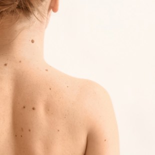
During your mole mapping appointment, we will take standardised photographs of your body and individual lesions. You will also be examined by a dermatology clinician using a dermatoscope (microscope) to examine individual lesions.
For the photographs you will be required to undress to your underwear, including removing your shoes and socks. The more skin that is exposed for the photographs the more lesions the device is able to image. However, it is very important that we respect your dignity at all times. For female patients, removal of the bra during photographs or clinical assessment is a personal choice and will depend on the number of lesions in this area. Likewise, individual lesions which are covered on the buttocks or in the genital area can be examined if required; although we do not take images in the genital area.
The mole mapping device takes standardised images which requires you to stand in specific poses and positions to ensure that the images are consistent for comparison at your next mole mapping appointment. We will talk you through these movements as the device takes the necessary images.
The whole process of mapping and examination will take approximately half an hour. If there are multiple lesions to image and assess it may take longer.
It is very important that you feel comfortable throughout the mole mapping process, please ask questions before or during your appointment if you are at all unclear or hesitant about anything.
Dermoscopy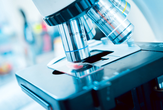
Use of a dermatoscope (looking at moles under a microscope) significantly increases the ability of a dermatologist to assess your skin lesions. Subtle changes in lesion structures, such as pigment and vessels can indicate concerning change. Likewise, a lack of such features can provide reassurance.
At The Private Medical Group all mole mapping is undertaken in conjunction with dermoscopy, performed by a doctor with specialist training in this area.
The combination of both mole mapping and dermoscopy by a trained clinician is the best way to keep track of changes in your skin lesions and identify those which may be of concern.